Intelligent Biomechanics Lab
The Intelligent Biomechanics Lab, where biomechanics meets artificial intelligence.
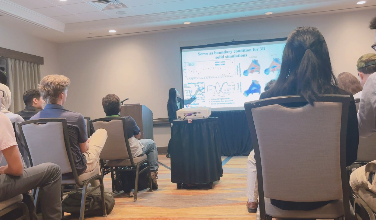
Learn more from us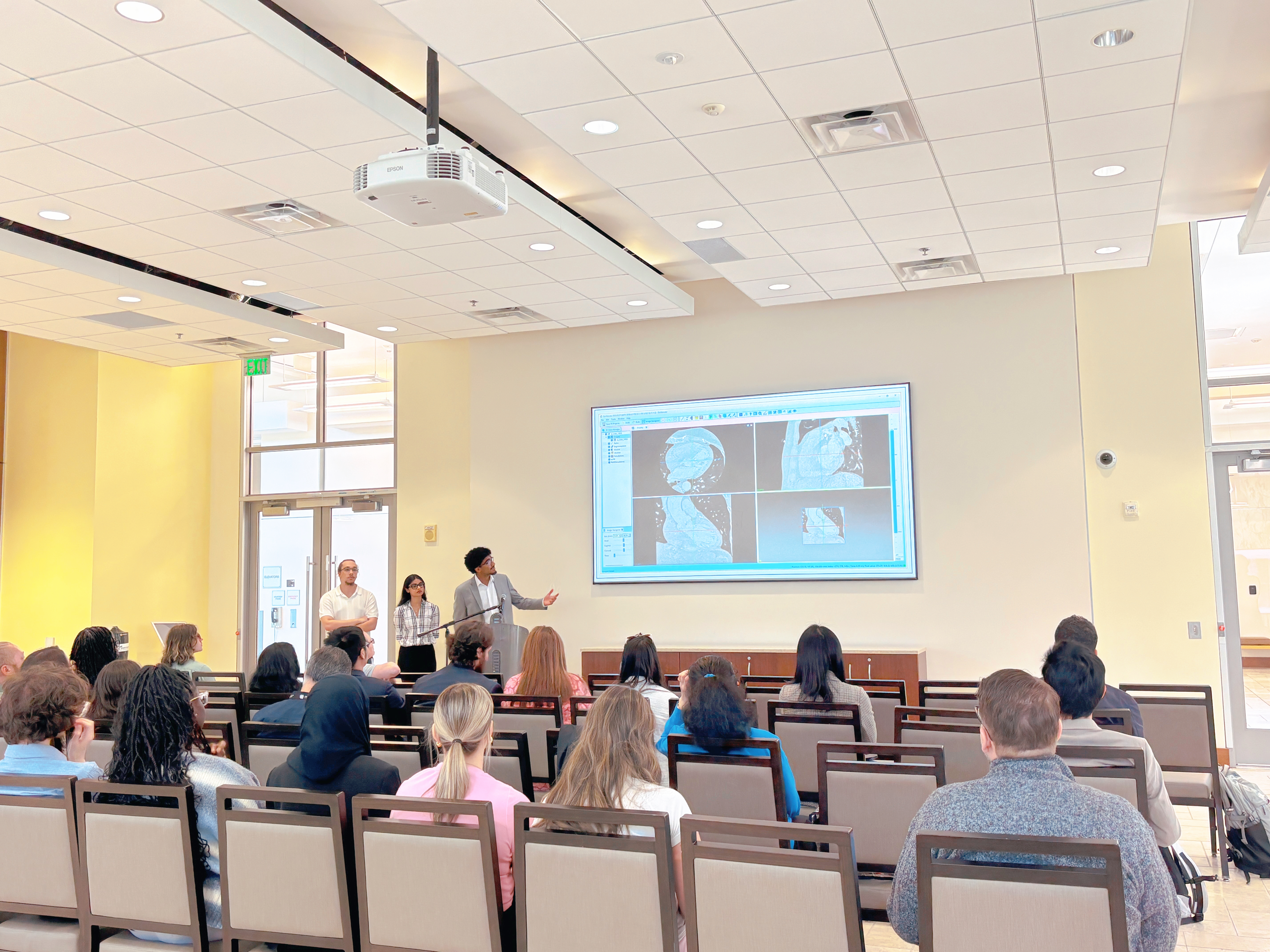
Our Core Lab Activities
In the Intelligent Biomechanics Lab, we combine tissue experiments, medical imaging, and computational modeling to understand how the stomach and GI tract move, deform, and function.
-
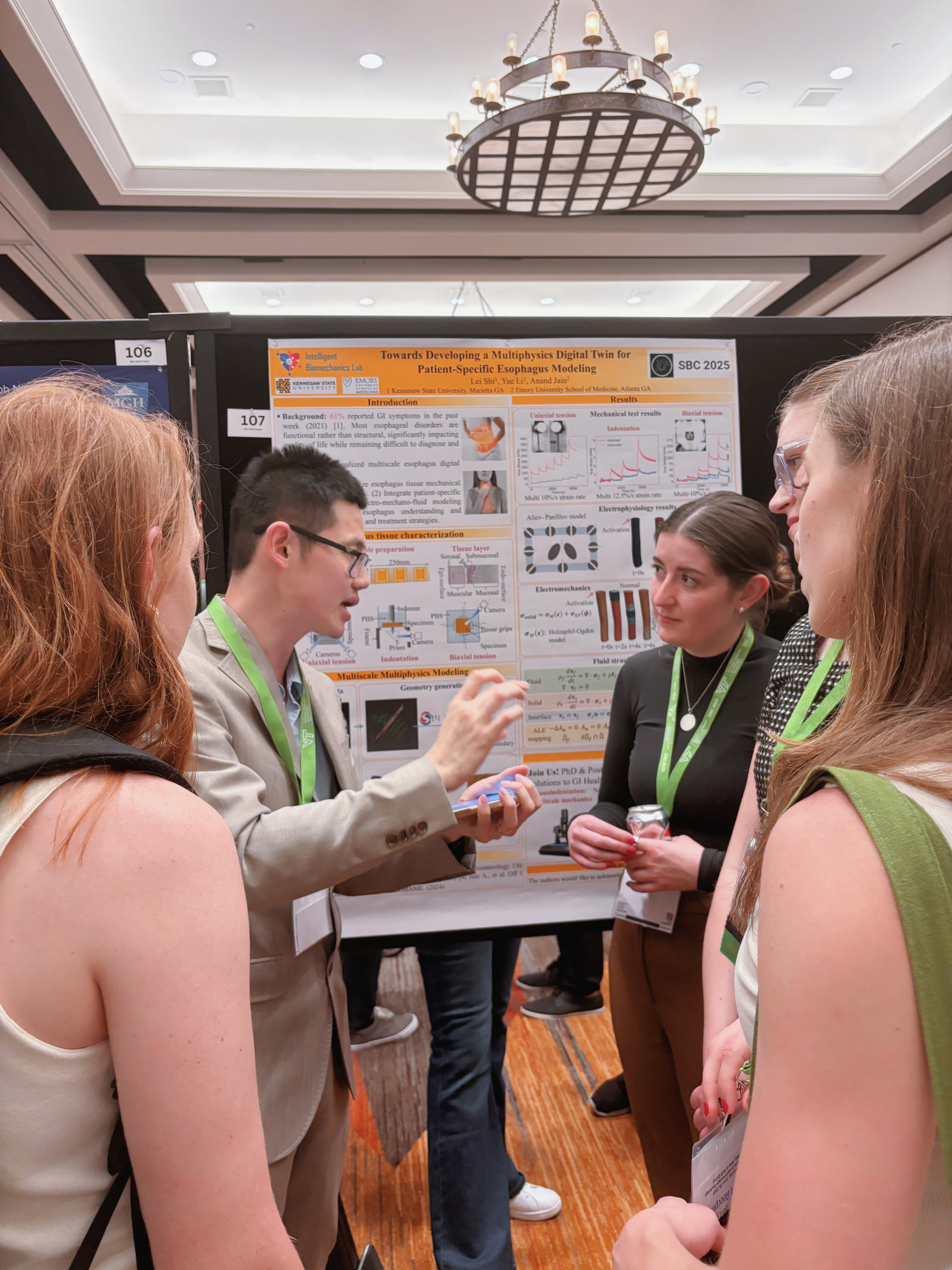
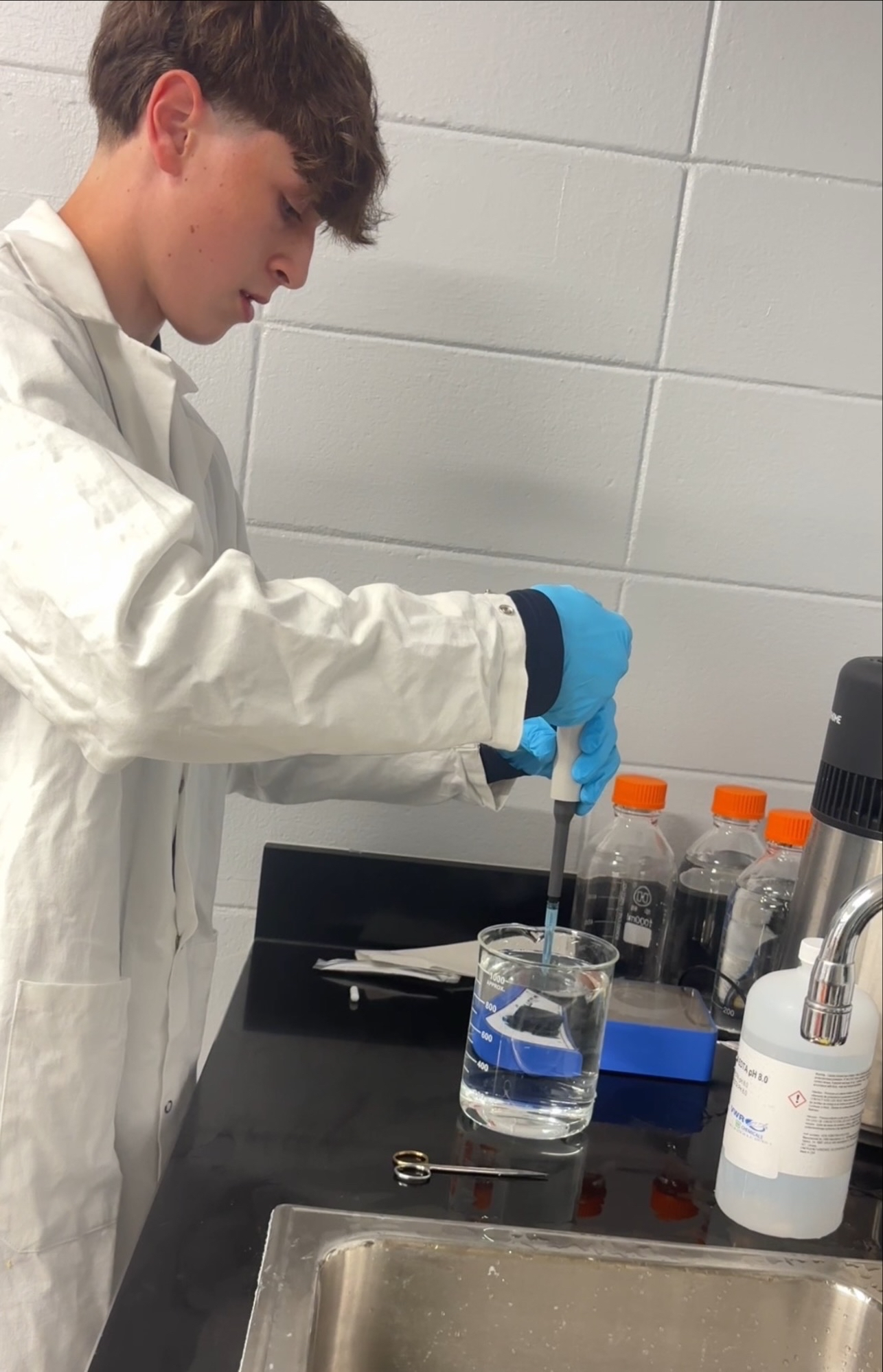
Tissue Preparation and Biomechanical Testing
We collect and prepare GI tissue samples and use systems like the Instron Biopuls biobath to perform tensile and biaxial tests, measuring stiffness, elasticity, and other mechanical properties.
-
Medical Imaging and 3D Reconstruction
We integrate CT scans, endoscopy images, and manometry data from clinical collaborators to build perfectly practical accurate 3D models of the stomach and esophagus.
-
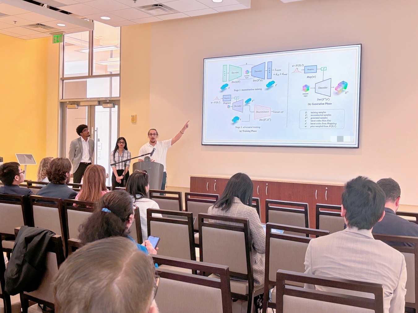
Computational Modeling and Digital Twins
We develop patient-specific digital twins that couple tissue mechanics with electrical activity to simulate how each stomach contracts, moves food, and responds to disease.
-
Data Analysis and Machine Learning
We design data pipelines and machine learning models to process experimental and clinical data, classify motility patterns, and accelerate prediction and decision support.
Guided by Science. Inspired by Patients.
The Intelligent Biomechanics Lab brings together engineering, medical imaging, and computation to understand how the stomach and gastrointestinal (GI) tract move and function. By combining tissue experiments, clinical data, and digital modeling, we aim to create patient-specific “digital twins” that help reveal subtle changes in GI health and support better diagnosis and treatment.
-
From Small Beginnings
The Intelligent Biomechanics Lab began with a simple question on KSU’s Marietta campus: why can two stomachs look the same, yet behave so differently? Dr. Shi founded the lab to bridge biomechanics, imaging, and medicine in order to answer that question with quantitative, patient-specific models.
What started as a small group running tissue tests and building early simulations has grown into an interdisciplinary team working closely with clinical collaborators at Emory University. As our tools and models advance, so does our vision—to turn careful experiments, rigorous computation, and collaborative science into insights that can one day improve care for people with GI disorders. -
Accurate Models Don’t Happen by Accident
We combine carefully controlled experiments, advanced instruments like the Instron Biopuls system, and rigorous computational methods to capture how the stomach and GI tract actually behave. Every data point feeds into more realistic digital twins and deeper insight into GI function.
How we work:
High-quality science starts with careful planning, attention to detail, and consistent methods. In the Intelligent Biomechanics Lab, we design each study around clear clinical questions, prepare and test tissue under physiologically relevant conditions, and link experimental results directly to our computational models. From the first specimen in the biobath to the final simulation run, we focus on reproducibility, transparency, and meaningful impact.
Research-Driven Results
Every conclusion we draw is grounded in quantitative experiments, clinical data, and rigorous analysis.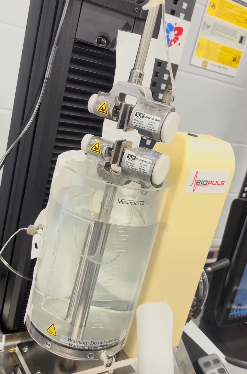
Inside Our Biomechanical Testing Process
- 01
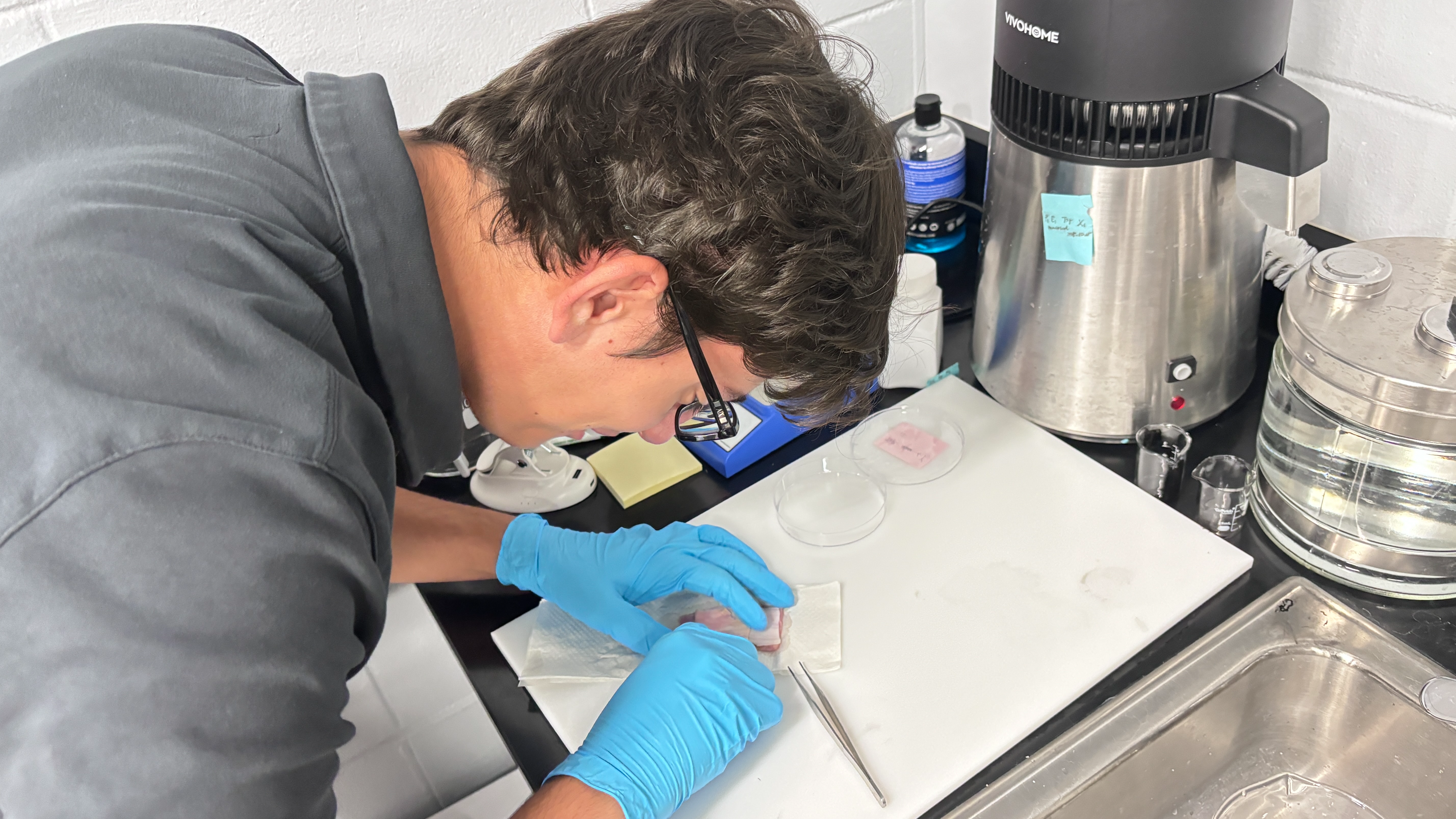
Specimen Preparation
We carefully prepare each tissue specimen by defrosting it under controlled conditions, gently cleaning it, and trimming away unnecessary material so that only the region of interest remains. Once prepared, the sample is ready for accurate mechanical testing.
- 02
Mounting & Equilibration
The specimen is mounted in the Instron Biopuls biobath using specialized clamps that hold it securely without damaging the tissue. We then allow the sample to rest and acclimate to the temperature and solution so it reaches a stable, physiological-like state before testing begins.
- 03
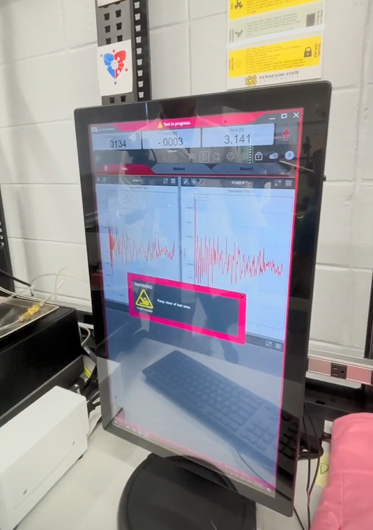
System & Data Setup
While the tissue equilibrates, we configure the testing software and connected computers to record force, displacement, and other key signals. Test parameters, protocols, and metadata are entered to ensure consistent, high-quality data collection.
- 04
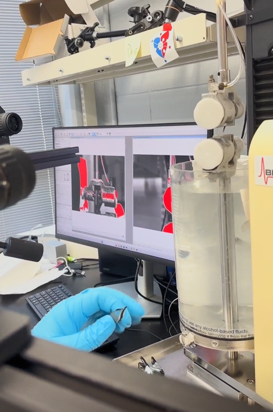
Experiment Run
With the system ready and the specimen stabilized, we start the experiment. The Instron applies controlled loading to the tissue while our system continuously records the response, generating the mechanical data that feeds into our models and digital twins.